Patellofemoral Implants
The Arthrosurface Patellofemoral Arthroplasty Systems utilize the inherent advantage of its proprietary Inlay Technology to address the full treatment spectrum of patellofemoral pathology including:
- Isolated PF Arthrosis and Arthritis
- PF Malalignment
- PF Instability
- Dysplasia
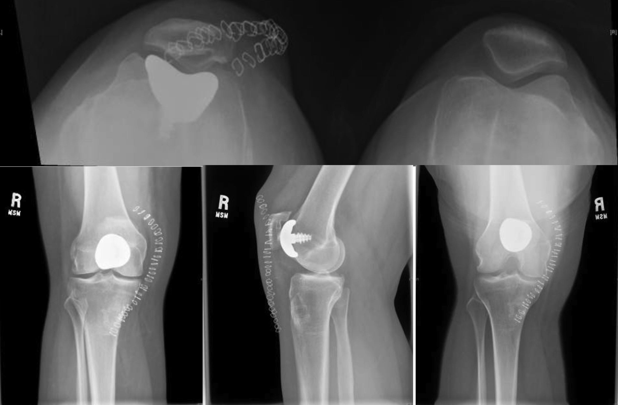
The Patellofemoral (PF) Implant Systems (PF Classic HemiCAP®, PF Wave® & WaveKahuna™) are designed to restore the unique articular surface geometry of the Patella and Femoral Trochlea Groove while maintaining patients’ native joint anatomy. This creates a congruent pathway by using an intraoperative 3-dimensional mapping system with contoured articular resurfacing implants. The PF Classic HemiCAP®, PF Wave® & WaveKahuna™ are available in 3 different sizes, with 29 trochlear convexities and 16 different patellas, making it the most versatile PF system in the world. The PF Classic system is indicated for isolated and contained lesions of the trochlea groove and patella. The PF Wave® and WaveKahuna™ Arthroplasty Systems are indicated for those patients who have more diffuse and/or extensive damage to their PF joint. Both the trochlea and patella implants are designed as inlay implants in order to maintain and restore congruency in the PF joint which is very important for proper patella tracking.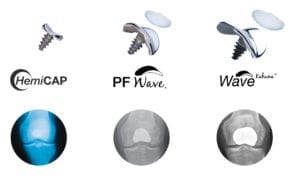
A recent study showed, “None of the patients in the inlay group showed progression of tibiofemoral OA. In contrast, the onlay group showed significant progression… with 53% of the
patients showing progression of medial and/or lateral tibiofemoral OA.” Followed by a study at 5 years with the same results, “At 5 year follow-up…no significant progression of tibiofemoral OA was observed.”
- Preserves native anatomy and can be performed in conjunction with soft tissue alignments and/or on dysplastic femurs
- Precision milling jigs provide a reproducible technique that eliminates repetitive trialing and burring
- Intraoperative mapping, multiple convexities and trials ensure an anatomic fit
- One tray with simple reproducible technique and instrumentation
- Multiple inlay trochlea and patella implants provide an off-the-shelf custom fit
- Inlay components preserve the joint by only removing a small amount of bone which maintains future options and native anatomy: “no bridges burned” should a future revision be needed.
- No activity restrictions following recovery.
- Can be performed as an outpatient surgery
- Slows progression of osteoarthritis
The fit achieved using the precision instrumentation of either the UniCAP® or PF HemiCAP® system is a significant improvement over existing joint replacement products. Arthrosurface® fits the implant to the patient at the time of surgery. Arthrosurface® restores the native joint biomechanics and preserves bone and tissue that is typically removed with total joint replacement systems. By leaving all supporting tissues intact, proprioception is maintained. Our goal with the UniCAP® or PF HemiCAP® system is to restore normal joint function by preserving native anatomy and resurfacing only the damaged surfaces in the knee.
The PF HemiCAP® technology was designed so patients can continue working and retain an active lifestyle without compromising future treatments.
- The anatomic femoral curvatures and inlay patella are designed to restore a natural moving knee by keeping the joint congruent and ligament structures intact
- The procedure may be performed on outpatient basis
- PF Systems allows for preservation of bone and soft tissues
- Maintains existing joint biomechanics thereby allowing normal motion and less recovery time
- Patients are experiencing a rapid return to activity for both work and exercise
- Shortener recovery time
- No activity restrictions
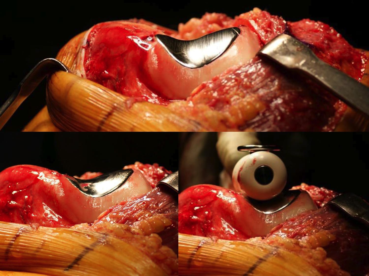
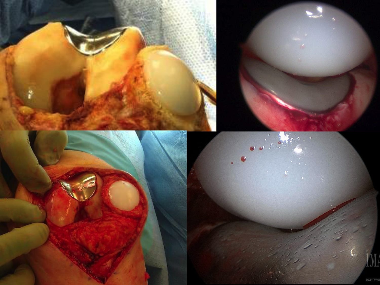
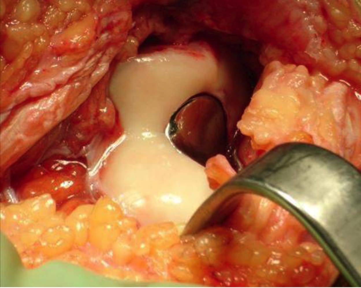
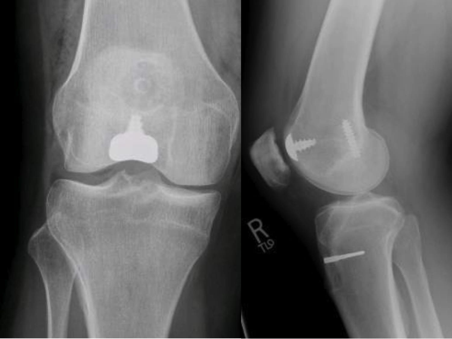
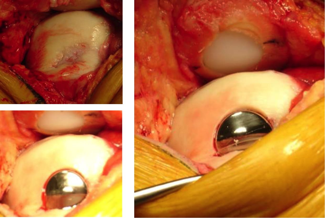
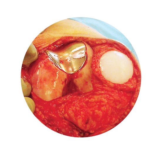
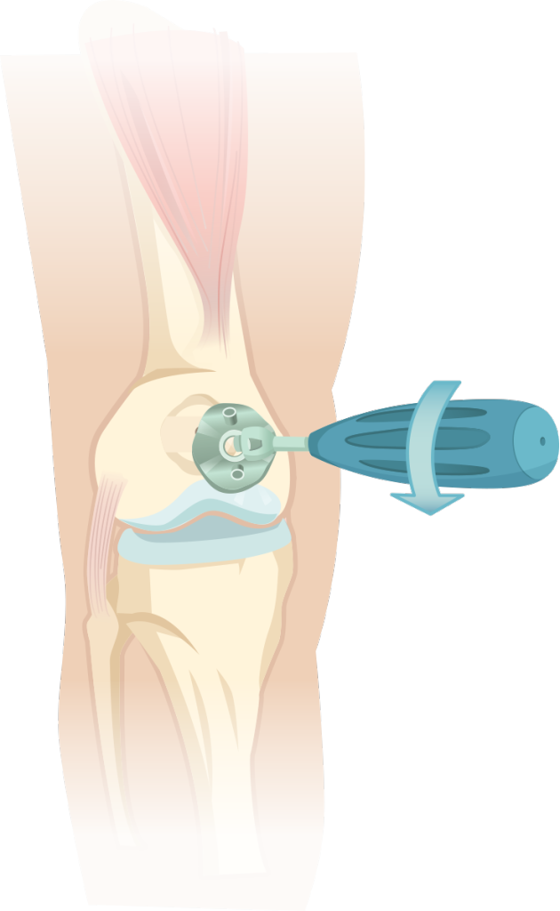 The Patello Femoral HemiCAP® implants are comprised of a two part system, the articular cap and a fixation component, as well as a cemented poly patella. The instruments are designed and organized in the order of surgery, proceeding from left to right and top to bottom. Resurface the femur prior to preparing the patella. Templates are used to determine the right surface curvature for the trochlea and then a jig is used to create 3 overlapping inlay sockets. The screw is then prepared through the trial in order to set it perpendicular to the articular implant. Once the recessed fit is confirmed, proceed with the patella procedure. A guide-wire is placed centrally, the socket reamed to the appropriate depth and then trials are used to confirm the best fit. Cement is placed in the patella socket and on the femoral component with both implants placed and set into the reamed sockets.
Intuitive, universal, reproducible procedures across multiple joints
- Minimally invasive procedure can be performed on an outpatient basis
- Ligaments or a realignment procedure can be performed in conjunction with the implant
- Maintains soft-tissue envelope and native joint mechanics
- Preserves meniscus and skeletal anatomy allowing a future total joint replacement
- The two components are connected together via morse taper, with zero reported loosening
- Less OR time with this quick, reproducible procedure